
 05
05


05

04

02

10

06
05

04
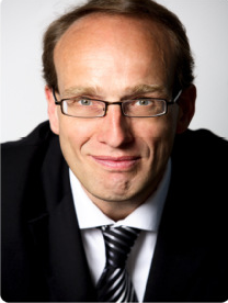
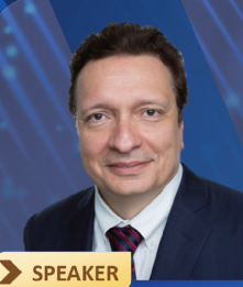
 Speaker:Georg Northoff
 Institution:University of Ottawa
 Time:28th May,14:30AM
 Locatiom:Zhonglan Meeting Room
Speaker: Li Zhang
Institution:University of Southern California
Time:26th May, 9:30AM
Locatiom:Meeting Room 705
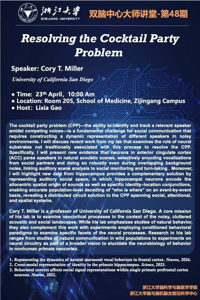
Speaker: Cory T. Miller
Institution:University of California San Diego
Time:23th April, 10:00 Am
Locatiom:Room 205, School of Medicine
Speaker:Chen Tao
Institution:Air Force Medical University of PLA
Time:11th December,10:00 AM
Locatiom:Meeting Room 705
Speaker:Julio Licino
Institution:Medical University
Time: 14th October,15:00 PM
Locatiom: Liangzhu Laboratory

Speaker:WANG Yutian
Institution:Fudan University
Time:13th October,10:00 AM
Locatiom:Liangzhu Laboratory

The School of Brain Science and Brain Medicine, devoted to the study of neuroscience and neuromedicine, was founded in October 2019. As the first school focusing on brain science and brain medicine in Chin... 【More】


Address : Zijingang Campus of Zhejiang University
Tel : 0571-87071107
E-mail : brains@zju.edu.cn